The goal of this project is to accurately segment the left atrium from MRI images, providing clear boundaries for better clinical assessments. The segmentation is achieved using a robust U-Net model, tailored for biomedical image segmentation tasks.
The U-Net model is specifically designed for biomedical image segmentation tasks. Its architecture consists of an encoder-decoder structure with skip connections, ensuring the preservation of spatial information.

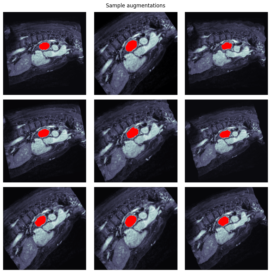
The training process involves feeding preprocessed MRI slices into the U-Net model with corresponding segmentation masks. Data augmentation techniques improve the model’s robustness to variations in input data.

Click on the image below to download and watch a sample MRI tested using the model

Atrium-Segmentation/ ├── data/ # Raw MRI images and labels ├── Preprocessed/ # Processed images and masks ├── models/ # Trained model weights ├── utils/ # Helper functions for data handling ├── notebooks/ # Jupyter notebooks for EDA and model training ├── Atrium Segmentation Evaluated on a subject.mp4 ├── Atrium Segmentation Training.png ├── unet.png └── README.md # Project documentation
The U-Net model achieves:
This result demonstrates the model’s strong ability to segment atrial structures accurately.
Contributions are welcome! To contribute:
1. **Fork** the repository.
2. **Create a new branch:**
git checkout -b feature-branch
This project is licensed under the MIT License. See the LICENSE file for details.
Special thanks to:
The medical imaging community for datasets and resources. U-Net developers for the foundational architecture. nibabel for neuroimaging data processing.